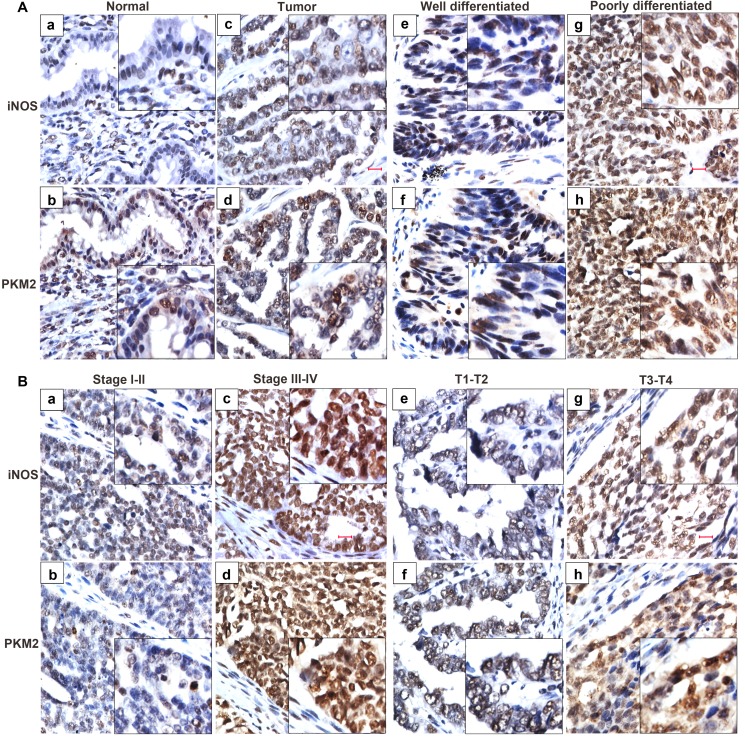
Figure 7. iNOS expression predicts an aggressive phenotype of ovarian cancer specimens.
The expression of iNOS and PKM2 is evaluated in 150 ovarian cancer tissues and 10 normal ovarian epithelial tissues by immunohistochemistry method. (A) iNOS and PKM2 staining in ovarian cancer tissues (c, d) is stronger than that in the normal ovarian epithelial tissues (a, b). The expression of iNOS and PKM2 were strongly stained in the poorly differentiated (g, h), while weakly stained in well differentiated ovarian cancer (e, f). (B) Representative images of iNOS and PKM2 in ovarian cancer tissues of different TNM stages. The expression of iNOS and PKM2 were strongly stained in the stage III-IV (c, d) and T3-T4 (g, h), while weakly stained in stage I-II (a, b) and T1-T2 (e, f) of cancer tissues.

