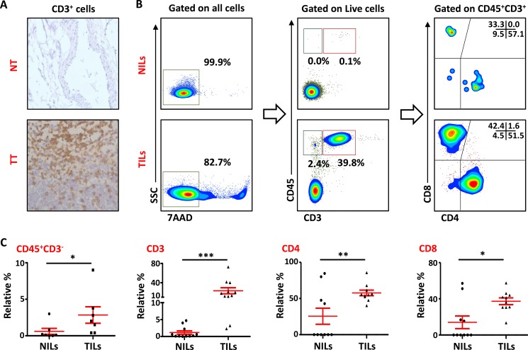
Figure 1. T-cell infiltration in normal and tumor tissues in primary breast cancer.
(A). Representative images of immunohistochemical staining of tumor-infiltrating CD3+ T cells in formalin-fixed paraffin embedded breast non-tumor (NT) and tumor tissues (TT). (B). Freshly isolated immune cells infiltrating NT (NILs) and TT (TILs) from 11 PBC patients were stained with 7AAD, CD45, CD3, CD4 and CD8 antibodies for identification of T cells and their subsets. Representative flow cytometric plots of surface staining from one cancer patient are shown. 7AAD dye was used to gate live cells, followed by lymphocyte identification by CD45 and CD3 stainings. Different subsets of T cells were then characterized using CD4 and CD8 antibodies. (C). Scatter plots showing the differences in tissue-infiltrating CD45+CD3−, CD45+CD3+, CD4+ and CD8+ cells between NILs and TILs.

