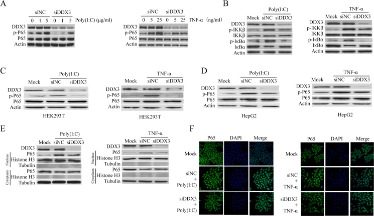
Figure 2. DDX3 regulates NF-κB signal pathway.
HeLa cells were first transfected with siDDX3 for 48 hours to silence DDX3 expression before the phosphorylation of p65 was tested by western blot after the cells were either transfected with Poly(I:C)(1 μg/ml or 5 μg/ml) for 6 hours or stimulated with TNF-α (5 ng/ml or 25 ng/ml) for 6 hours (A). The same experiment was also performed in HEK-293T (C) and HepG2 cells (D). The phosphorylation of IκBα and IKK-β, as well as the degradation of IκBα, were also determined after the cells were treated by 5 μg/ml poly(I:C) or 25 ng/ml TNF-α (B). The level of p65 in the cytoplasm and nucleus were detected after HeLa cells were stimulated with poly(I:C) or TNF-α (E). For IFA detection, HeLa cells were first seeded on glass cover slips for 24 hours before transfected with siDDX3 for 48 hours to silence DDX3 expression. After stimulated with poly(I:C) (5 μg/ml) or TNF-α (25 ng/ml) for 1 hour, the cells were fixed and immunostained for p65 using a rabbit anti-p65 antibody and Alexa Fluor® 488 goat anti-rabbit IgG (H+L) secondary antibody. Nuclei were counterstained with DAPI. The images were captured by confocal microscopy (F). The scrambled siRNAs (siNC) were used as the control.

