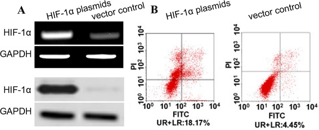
Figure 5. HIF-1α induces apoptosis of aortic SMCs.
(A) Aortic SMCs were infected with HIF-1α plasmids (or vectors) at a MOI of 100. The expression of HIF-1α and its effect on cell apoptosis were detected at 72 hr post-infection. The expression level of HIF-1α mRNA and protein was detected by qRT-PCR and western blot and normalized to that of GAPDH. (B) Cells were stained with annexin V-FITC and propidium iodide (PI). Flow cytometric contour plots showed the percentage of stained cells. Histogram showed the percentage of Annexin V+/PI- and Annexin V+/PI+ cells of HIF-1α-infected and vector-infected aortic SMCs.

