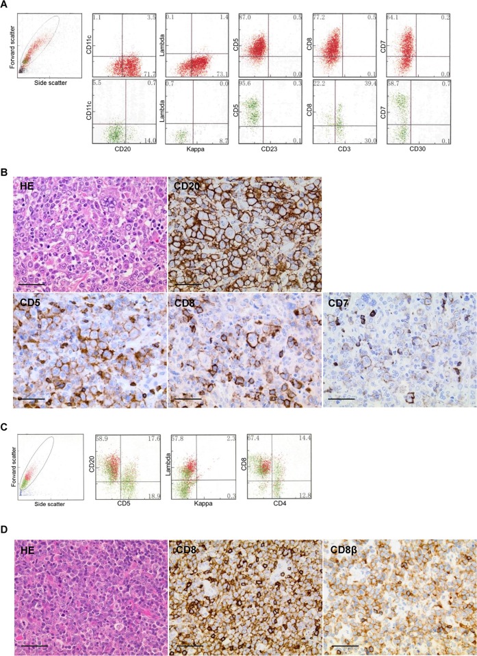
Figure 2. Morphological and immunophenotypic features of T-cell marker-positive diffuse large B-cell lymphomas.
A and B, case 1; C and D, case 16. Scale bars represent 50 μm. (A) Flow cytometry (FCM) showed an abnormal large cell population (red colored plots) positive for CD20, kappa chain, CD5, CD8, and CD7, with a broad range of fluorescent intensity from cells negative or dimly positive for CD7. (B) Histologically, lymphoma cells showed a pleomorphic large cell morphology containing multinucleated giant cells (hematoxylin and eosin stain, HE; ×40). Immunohistochemistry showed strongly positive diffuse staining for CD20, weakly positive staining for CD5, and focally positive staining for CD8 and CD7. (C) FCM showed the formation of clusters, with positive staining for CD20, lambda chain, and CD8 (red colored plots). The intensity of CD8 was as strong as that of the background normal T cells (green colored plots). (D) The cell morphology showed centroblastic large cell infiltrates (HE, ×60). Immunohistochemically, the majority of the lymphoma cells were positive for both CD8 and CD8β. Note that the positive staining of T-cell markers on tumor cells was weaker than on admixed normal T cells, in both case 1 and case 16.

