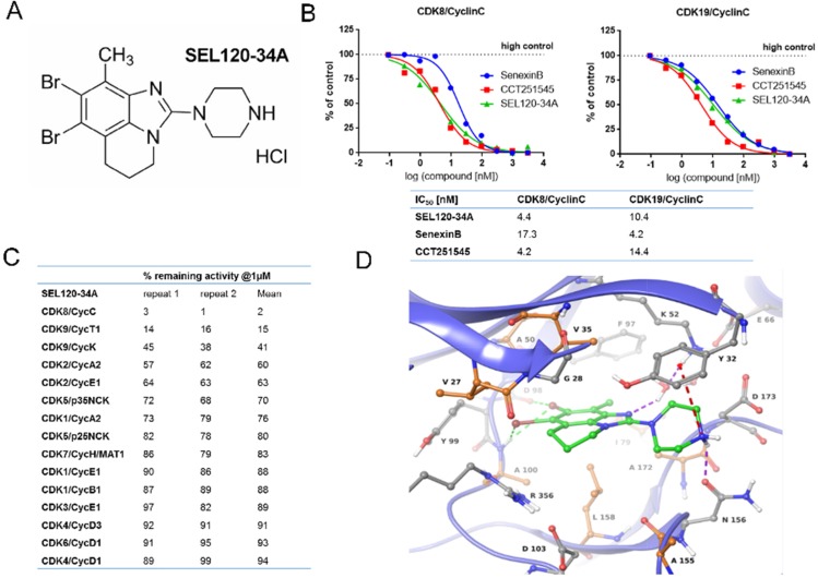
Figure 1. Structure and activity of SEL120-34A.
(A) Chemical structure of SEL120-34A. (B) The IC50 of SEL120-34A, Senexin B and CCT241545 determined by constructing a dose-response curve and examining inhibition of CDK8/CycC and CDK19/CycC activities at Km ATP concentrations. (C) % remaining activities measured for members of the CDK family in the presence of 1 μM SEL120-34A at Km ATP concentrations. (D) Active site of the crystal structure of human CDK8/CycC complexed with SEL120-34A. Protein residues and SEL120-34A are shown as Ball-and-Sticks. Protein carbon atoms are colored orange (aliphatic hydrophobic residues) or gray (other residues), while ligand carbon atoms are colored green. The following interactions are shown: H bond as purple dashed line, halogen bonding as green dashed line and cation-π system interaction as red dashed line.

