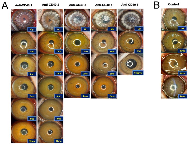
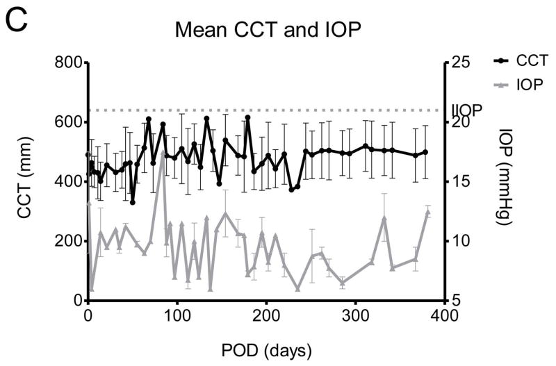
Figure 1. Representative photographs of grafted eyes showing clinical courses, and changes of central corneal thickness and intraocular pressure in deep lamellar porcine keratoplasty in nonhuman primates.
(A) All grafts in the anti-CD40 Abs treated primates survived at least 6 months except one which accidently died due to anesthetic event at POD 61. The primate also showed clear graft until death. Two of the grafted primates showed more than 1year survival. (B) Representative photos of the control showing rejection within 4 weeks. (C) Central corneal thickness was well maintained under 600 μm and increase of intraocular pressure was not observed during the follow-up.


