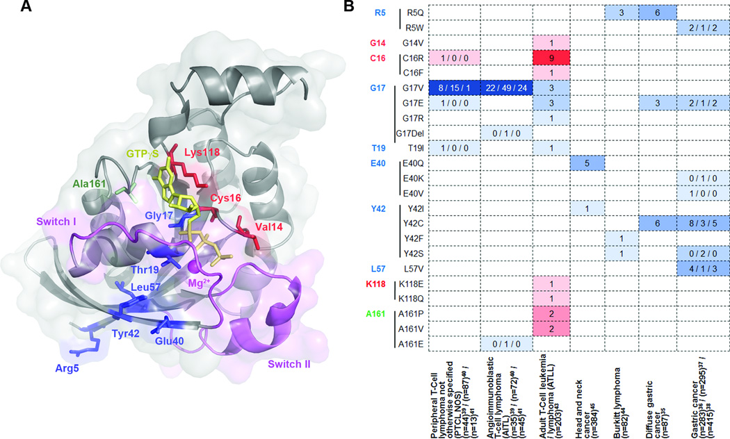
Figure 2.
Recurrent RhoA mutations found in human cancers. (a) Most commonly mutated amino acid positions identified in recent reports are mapped to the 3D structure of activated RhoA161 (PDB#: 1A2B). GTPγS is shown as yellow sticks and the magnesium ion is shown as magenta sphere. Sites for gain-of-function mutations G14 (shown as V14, mutated in the original structure), C16, and K118 are shown as red sticks; sites for loss-of-function mutations R5, G17, T19, E40, Y42, and L57 are shown as blue sticks; while A161 is shown as green sticks as both gain-of-function mutations (A161P and A161V) and lost-of-function mutations (A161E) have been identified. (b) The occurrence of the hot-spot mutations are listed across tumor types. Hotspots are ordered by amino acid position and colored in the same scheme as in (a). Gain-of-function and loss-of-function mutations are classified based on preliminary biochemical studies and need further characterization. In Palomero et al.39, RhoA mutations other than G17V were identified in a single case each and the authors didn’t specify the PTCL subtype. They are included in PTCL-NOS here for simplicity.

