Figure 2.

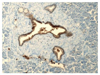
Benign pancreatic ducts and acini with weak staining of the ductal cells for carcinoembryonic antigen (200 ×). Courtesy, Department of Pathology, Temple University Hospital, Philadelphia, PA, United States.

Benign pancreatic ducts and acini with weak staining of the ductal cells for carcinoembryonic antigen (200 ×). Courtesy, Department of Pathology, Temple University Hospital, Philadelphia, PA, United States.