
Figure 2.
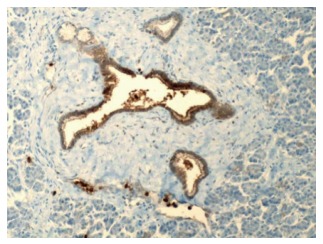
Benign pancreatic ducts and acini with weak staining of the ductal cells for carcinoembryonic antigen (200 ×). Courtesy, Department of Pathology, Temple University Hospital, Philadelphia, PA, United States.
Official websites use .gov
A
.gov website belongs to an official
government organization in the United States.
Secure .gov websites use HTTPS
A lock (
) or https:// means you've safely
connected to the .gov website. Share sensitive
information only on official, secure websites.
Benign pancreatic ducts and acini with weak staining of the ductal cells for carcinoembryonic antigen (200 ×). Courtesy, Department of Pathology, Temple University Hospital, Philadelphia, PA, United States.