Figure 6.

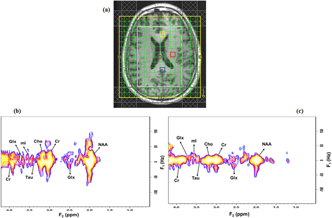
(a) T 1-weighted axial MRI of a 59-year-old healthy brain with the white box indicating the PRESS localization, (b) selected 2D J-resolved spectra extracted from the mid occipital (voxel in blue) and, (c) mid frontal (voxel in yellow).

(a) T 1-weighted axial MRI of a 59-year-old healthy brain with the white box indicating the PRESS localization, (b) selected 2D J-resolved spectra extracted from the mid occipital (voxel in blue) and, (c) mid frontal (voxel in yellow).