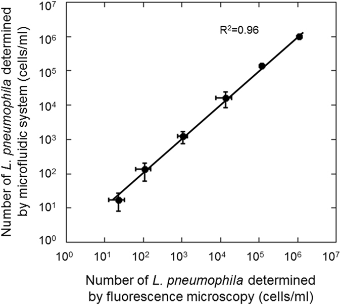
Figure 3.

Correlation between microfluidic counts and conventional fluorescence microscopic counts of L. pneumophila stained with a fluorescent antibody. Cultured L. pneumophila cells were spiked in cooling tower water. Error bars indicate the standard deviation (n = 5). Samples with 101 cells/ml of Legionella cells were counted following 1000-fold concentration by filtration. Samples with 102 and 103 cells/ml of Legionella were 100-fold concentrated before counting, and samples with 104 to 106 cells/ml of Legionella were counted without concentration.
