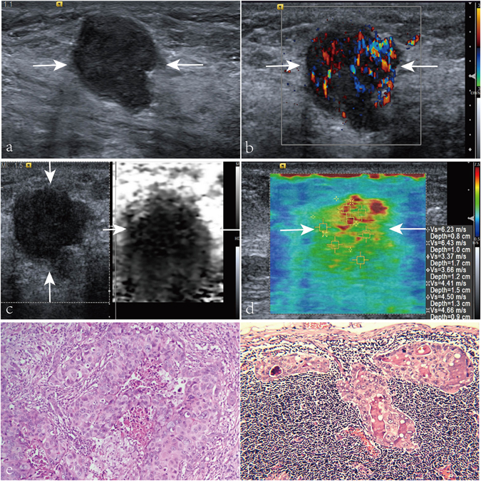
Figure 2.

Images in a 58-year-old patient with breast invasive ductal carcinoma, axillary lymph node metastasis (LNM), histologic grade III, negative estrogen receptor (ER), negative progesterone receptor (PR), and positive C-erbB-2. (a) A solid, marked hypoechogenicity, well defined margin, irregular, and taller than wide shape lesion (arrows) is shown on US. (b) Rich internal flow (i.e. 3 linear or tree-like signals) is found on color Doppler flow image (arrows) of the breast invasive ductal carcinoma. (c) Virtual touch tissue imaging (VTI) score of the lesion (arrows) is 4. (d) On virtual touch tissue imaging & quantification image, the lesion (arrows) is heterogeneous with a mean SWS value of 4.75 m/s. (e) Pathological examination confirms the diagnosis of invasive ductal carcinoma (Hematoxylin-eosin stain, ×200). (f) Pathological examination confirms the diagnosis of axillary LNM (Hematoxylin-eosin stain, ×200).
