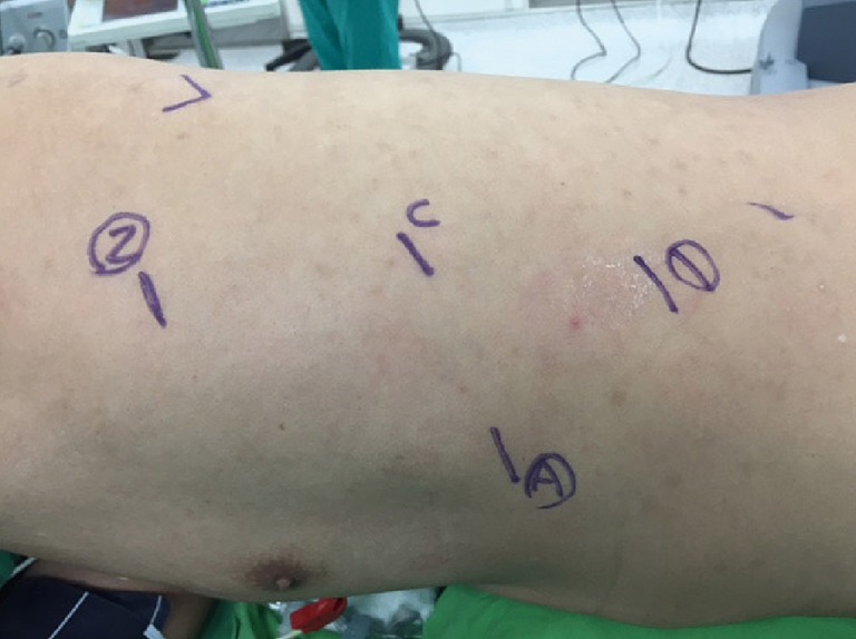
Figure 3.

Under general anesthesia with a double-lumen endotracheal tube, the patient placed in the right lateral semi-prone position. Incisions made at the 5th, 7th, and 9th intercostal spaces (ICSs). The left pleural space entered via dissection and a 12-mm trocar placed at the 7th ICS for the camera. The other 8-mm ports for the robotic arms and another assistant port placed at the 7th ICS, the anterior axillary line.
