
Figure 7.
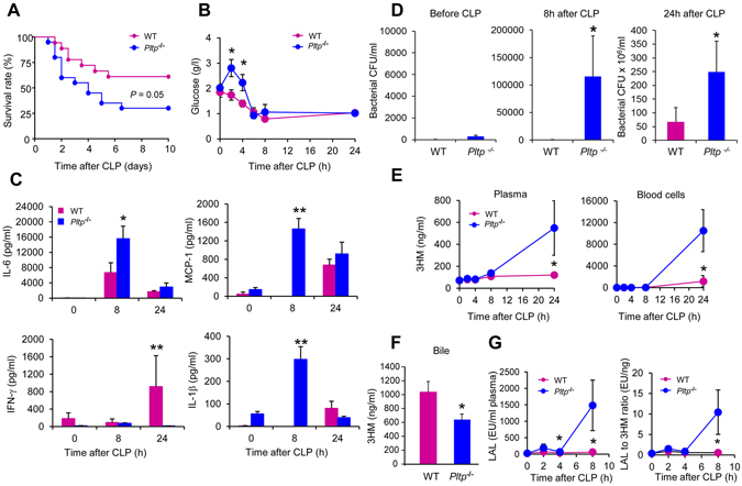
Endogenous PLTP expression protected mice against inflammation and death associated with polymicrobial sepsis. (A) PLTP deficiency increased mortality. Age-matched WT and Pltp−/− male mice underwent CLP. Data were analyzed using the Kaplan-Meier method, with statistical significance determined using the log rank test (n = 18–20 mice per group). (B) PLTP deficiency induced hyperglycaemia post-CLP. Blood glucose concentration was determined with a glucose meter before and at 2 h, 4 h, 6 h, 8 h and 24 h following CLP (n = 5 mice per group, t test). (C) PLTP deficiency increased the production of the inflammatory cytokines IL-6, MCP-1, and IL-1β and decreased that of IFN-γ. Plasma samples, harvested from WT and Pltp−/− mice before CLP, and 8 h and 24 h after CLP, were assayed using a Milliplex mouse cytokine panel (n = 5 mice per group, Mann Whitney test). (D) PLTP expression prevented sepsis by decreasing bacterial burden. Colony-forming units (CFU) were determined in blood samples harvested from WT and Pltp−/− mice before CLP, 8 h and 24 h following CLP (n = 11–13 mice per group, Mann Whitney test). (E-G) PLTP expression protected mice against polymicrobial sepsis by limiting the concentration and biological activity of LPS in the bloodstream, by increasing its biliary excretion, resulting in a higher plasma LPS neutralizing capacity. LPS concentrations in plasma and blood cells (E) and in bile (F) harvested from WT and Pltp−/− mice were determined following CLP by direct quantitation of 3HM (plasma and blood cells n = 5–6 mice per group, Mann Whitney test; bile n = 12 mice per group, t test). The biological activity of LPS in plasma (G) was quantified by LAL assay and the LPS activity index was calculated as the LAL to 3HM ratio (n = 6–7 mice per group, Mann Whitney test). Data are means ± sem. *P < 0.05, **P < 0.01.