
Figure 1.
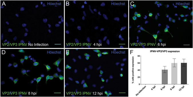
Timecourse of IPNV infection of CHSE-214 cells. VP2/VP3 expression was determined by immunofluorescence after 4 (B), 6 (C), 8 (D) and 12 (E) h post infection (hpi) in CHSE-214 cells. Cells were propagated at 60–70% confluence in 24-well plates with round glass coverslips. IPNV was inoculated at a MOI of 1 for 1 h. After 4, 6, 8 and 12 h of incubation at 20 °C, cells were processed by indirect immunofluorescence (IFI) using mouse oligoclonal anti-VP2/VP3 IPNV antibody and Alexa 488-conjugated donkey anti-mouse antibody. Representative images, recorded with an Olympus Spinning Disk IX81 microscope, are shown (bar scale = 20 μm). At each incubation time, 250 cells were counted. Number of VP2/VP3-expressing cells is shown as a percentage, normalized to mock cells (F).