
Figure 4.
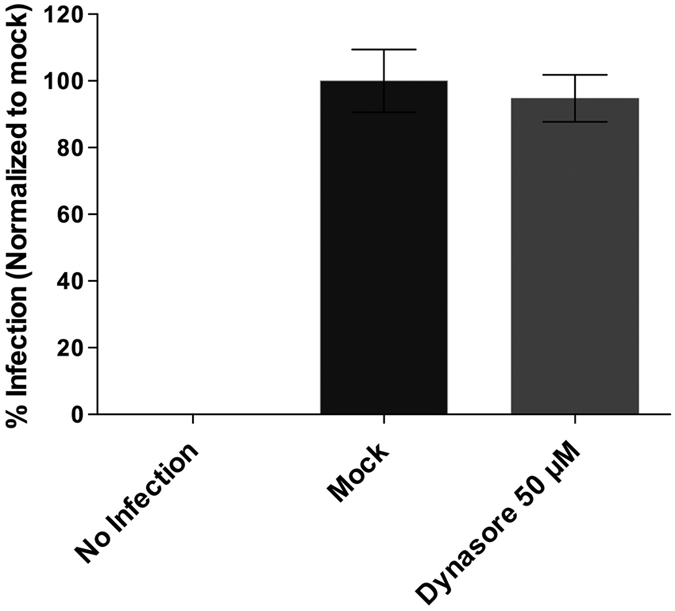
IPNV infection is independent of dynamin. CHSE-214 cells were propagated at 70–80% confluence in 24-well plates with round glass coverslips and pre incubated with 0 or 50 µM dynasore for 1 h at 20 °C. IPNV was inoculated at an MOI of 1 and led the infection proceed for 12 h at 20 °C in absence (mock) or presence of the inhibitor. Cells were processed by indirect immunofluorescence (IFI) for VP2/VP3 IPNV proteins. Imaging of fixed slides was performed with an Olympus Spinning Disk IX81 microscope, and 250 cells were counted at each condition. Number of VP2/VP3-expressing cells is shown as a percentage, normalized to mock cells.