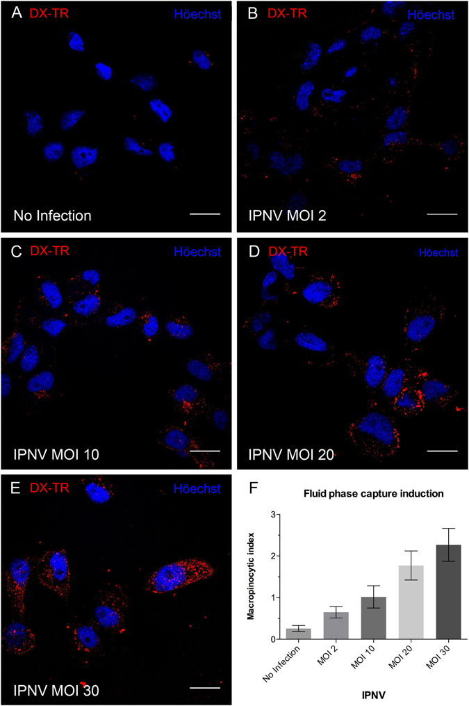
Figure 5.

IPNV induces fluid phase capture in CHSE-214 cells. CHSE-214 cells were grown to 70% confluence in 24-well plates with round coverslips in MEM with 2% FBS. Cells were left for 16 h in MEM without FBS. IPNV at MOI of 0, 2, 10, 20 and 30 (A–E) was inoculated, then left to adsorb for 1 h at 4 °C. Dextran Texas red (250 µg/ml) was added, and cells were incubated at 20 °C for 45 min. Cells were fixed and stained with Höechst. Images were recorded with a C2 Plus Eclipse TI Nikon confocal microscope. Representative images are shown (scale bar = 10 μm). A graph of the macropinocytic index vs MOI is presented in F. The macropinocytic index was determined as described in Materials and Methods.
