
Figure 6.
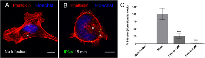
IPNV infection is dependent on actin polymerization dynamics. A. CHSE-214 cells were infected with IPNV at a MOI of 10 and adsorbed for 1 h at 4 °C. The supernatant was removed and MEM medium added at 20 °C. Cells were incubated at 20 °C for 15 min, fixed and stained with rhodamine-conjugated phalloidin. Images were recorded with a C2 Plus Nikon spectral confocal microscope, (scale = 10 μm). (A) no infection (B) 15 min post infection (white arrow heads indicate stress fibers in A and disorganized actin in B). (C) CHSE-214 cells were preincubated with 1 or 2 µM cytochalasin D (Cyto D) for 1 h and infected with IPNV at a MOI of 1 in the presence of Cyto D. Cells were incubated at 20 °C for 6 h and processed by indirect immunofluorescence (IFI) for IPNV VP2/VP3. For each condition, 250 cells were counted. The number of VP2/VP3-expressing cells is shown as a percentage, normalized to mock cells (****p < 0.0001).