Figure 3.

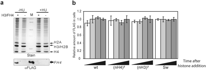
Exogenous histone complexes are stably incorporated into Physarum cells. (a) Hydroxy-urea treatment inhibits nuclear import. Cell fragments in early S-phase were untreted (−) and treated (+) with exogenous H3/FH4, and untreated (−HU) and treated with hydroxyl-urea (+HU), concomitantly. Nuclei were prepared and analyzed by SDS-PAGE (Stain) and Western blotting (αFLAG). Lane M corresponds to the molecular weight marker in (Stain) and the purified H3/FH4 complex revealed by anti-FLAG antibody in (αFLAG). (b) Determination of the stability of exogenous histone complexes in Physarum. Cell fragments were treated with HU and exogenous complexes were incorporated for 15 min, 30 min, 45 min and 60 min, respectively. Shown is the quantitative analysis of Flag signal relative to the amount of total soluble proteins determined by dot blotting. The quantification at time point 60 min was arbitrary assigned to 1.0 for each complex. Note that signals of untreated cell fragments with exogenous histones were ~10%. (c) Exogenous histone complexes are transported in nuclei with similar rates. The different histone complexes were spread onto Physarum surfaces and harvested after 20 min, 40 min and 60 min, respectively. Nuclei were then prepared and analyzed by Western blotting. Shown is the amount of exogenous histone complexes in the nuclear fractions at specific incorporation duration. The value 1 for each complex corresponded to the incorporation after 60 min.
