
Figure 2.
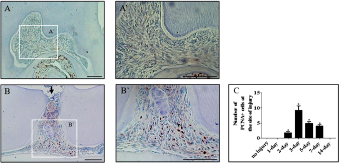
Pulp cells proliferate in response to damage. Immunohistochemical staining of proliferating cells with a PCNA antibody in an undamaged molar at low (A) and high magnification (A’). Immunohistochemical staining of proliferating cells with a PCNA antibody in a damaged superior first molar 3 days post-damage at low (B) and high magnification (B’). All damages were performed in CD-1 mice and representative sagittal sections are from four independent experiments. Scale bars are equivalent to 100 μm, an arrow indicates a pulp exposure. Quantification of the PCNA positive cells (C) was performed on three high-powered fields on at least four specimens per group, *p = <0.05.