
Figure 4.
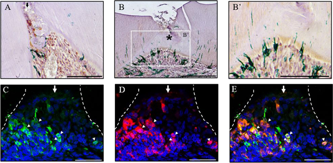
Odontoblast-like cells are descendants of Wnt active cells. Immunohistochemical staining of Axin2-expressing cells with GFP in a damaged superior first molar 3 days post-damage (A) and 14 days post-damage at low (B) and high magnification (B’). Immunofluorescent staining of Axin2-expressing cells with GFP (C), in situ hybridisation analysis of dspp expression (D) and merged image (E) of damaged superior first molar from Axin2CreERT2; Rosa26-mT-mG flox/+ mice 5 days post-damage. Representative sagittal sections are shown from four independent experiment’s. Scale bars are equivalent to 100 μm. The dentine-pulp interface is outlined by a white dashed line drawn from the light field image. An arrow indicates pulp exposure; arrow heads indicate examples of double stained cells and asterisk indicates the formation of a dentine bridge.