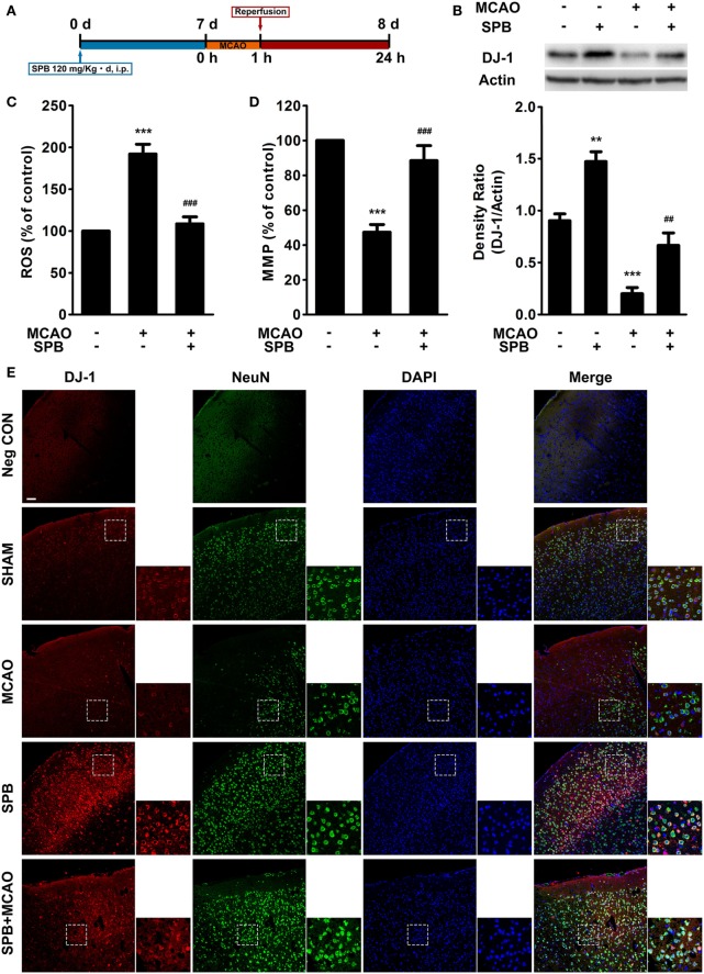
Figure 4.
Sodium phenylbutyrate (SPB) reverses the DJ-1 decrease and mitochondrial dysfunction induced by ischemia/reperfusion (I/R) injury in mice. (A) Experimental protocol of SPB treatment and the I/R model in mice. SPB was injected intraperitoneally at 120 mg/kg per day for 7 days. Middle cerebral artery occlusion (MCAO) was administered for 1 h on the eighth day as described in the methods, and then, reperfusion was established for 23 h. All of the subsequent animal experiments were performed 23 h after reperfusion unless otherwise stated. (B) Western blotting of DJ-1 in the mouse brain and quantitative analysis. The samples were from the peri-infarct region of MCAO mice or relative regions of sham surgery mice. **p < 0.01, ***p < 0.001, vs. the control group; ##p < 0.01, vs. MCAO without SPB treatment group, one-way analysis of variance (ANOVA), n = 6. Measurement of reactive oxygen species (ROS) production (C) and the mitochondrial membrane potential (D) in the peri-infarct region or relative regions of mice. ***p < 0.001, vs. the control group; ###p < 0.001, vs. MCAO without SPB treatment group, one-way ANOVA, n = 6. (E) Immunofluorescence analysis of DJ-1 and NeuN colocalization. The coronal sections were serially sectioned from the plane on which hippocampus begins to appear, and the cortex region were observed and displayed in the figure. The smaller pictures show the details in the boxed areas (red: DJ-1; green: NeuN; blue: DAPI; bar for 200 µm).

