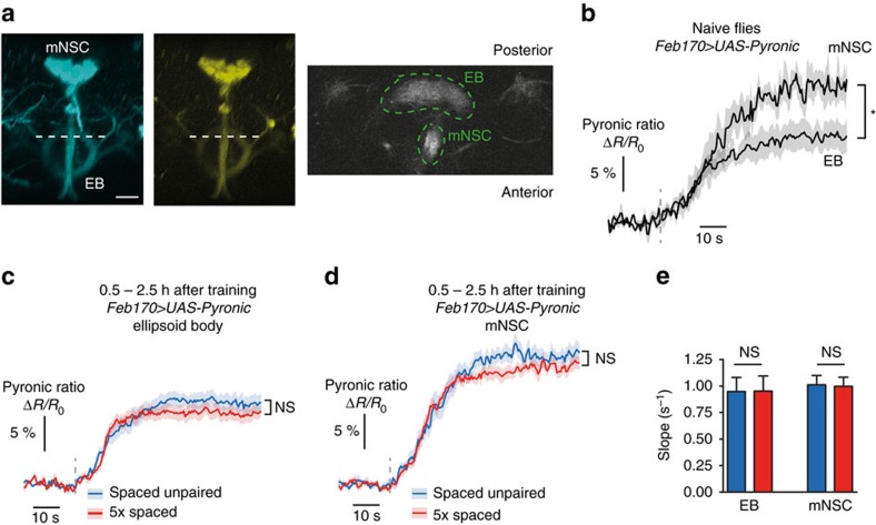
Figure 3. The energy flux in ellipsoid body neurons or in median neurosecretory cells is not increased following spaced training.
(a) Images of the Pyronic sensor expressed through the Feb170 driver, obtained by two-photon microscopy. The two images on the left show the 3D reconstruction of image stacks in the mTFP and Venus channels in the conditions that were used for live recordings (scale bar, 20 μm). This driver is commonly used to label ellipsoid body (EB) neurons, but it also strongly labels a cluster of ∼20 median neurosecretory cells (mNSC). Live recordings were obtained from a horizontal plane where both the upper part of the EB and a section of the descending branch from mNSC were visible (dashed white line). An illustrative example is shown on the right, with the corresponding regions of interest (green dashed lines). (b) Application of 5 mM sodium azide (grey dashed line) caused an increase in the Pyronic ratio in the EB and the mNSC (n=5). The plateau value was significantly lower in the EB than in the mNSC (t9=2.62; P=0.028). (c) Measurement of pyruvate accumulation in EB neurons following azide application after spaced training or control unpaired protocol. The plateau value was not statistically different between the two conditions (n=12–13; t23=1.47; P=0.15). (d) Measurement of pyruvate accumulation in mNSC following azide application after spaced training or control unpaired protocol. The plateau value was not statistically different between the two conditions (n=13; t24=1.37; P=0.18). (e) Average slope from data shown in c,d. The slope after spaced training was not different from after a spaced unpaired protocol in either structure (EB: t23=0.06; P=0.96; mNSC: t24=0.12; P=0.90).

