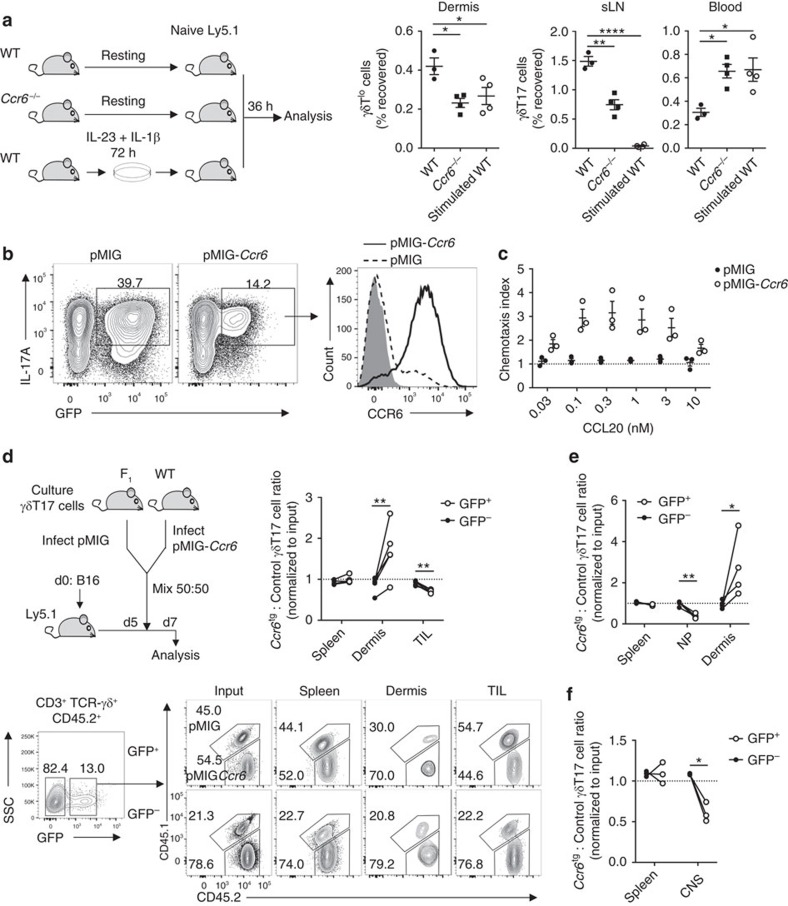
Figure 6. CCR6 downregulation by γδT17 cells enhances migration to inflamed tissue.
(a) Resting lymphocytes from wild type (WT) (n=3) or Ccr6−/− (n=4) mice, or WT lymphocytes stimulated with IL-23/IL-1β for 72 h (n=4) were transferred i.v. into separate naïve Ly5.1 hosts. After 36 h, number of CD45.2+ γδTlo/γδT17 cells recovered was expressed as % of number transferred. sLN, skin-draining lymph node. (b) Representative flow cytometry for CCR6 expression by GFP+ in vitro-expanded γδT17 cells transduced with empty pMIG or pMIG-Ccr6, relative to isotype (grey) (n=3). (c) Chemotaxis of GFP+ γδT17 cells transduced as in (b) to CCL20 (n=3). (d) In vitro-expanded γδT17 cells from F1 (CD45.1+CD45.2+) or WT (CD45.2+) mice were transduced with empty pMIG or pMIG-Ccr6, respectively. Equal numbers of mixed GFP+ cells were transferred i.v. into Ly5.1 mice challenged with B16 melanoma 5 days prior and analysed at d7 (n=5). Representative flow cytometry and ratio of recovered F1 to WT γδT17 cells within transduced (GFP+) and untransduced (GFP−) populations. Recovered values were normalized to input values. TIL, tumour-infiltrating lymphocytes. (e,f) In vitro-expanded γδT17 cells from WT or F1 mice were transduced with empty pMIG or pMIG-Ccr6, respectively. Equal numbers of mixed GFP+ cells were transferred i.v. into Ly5.1 mice either (e) 24 h post-infection with S. pneumoniae (n=4) or (f) at experimental autoimmune encephalomyelitis (EAE) onset (n=3) and organs were analysed 48 h later. Ratio of recovered WT to F1 γδT17 cells within transduced (GFP+) and untransduced (GFP−) populations, normalized to input values. CNS, central nervous system; NP, nasal passage. Mean±s.e.m. (a) Representative of three similar experiments, (b,d) representative of two experiments. (a) One-way ANOVA with Dunnett's multiple comparisons test relative to resting WT γδT17 cells, (d–f) paired two-tailed Student's t-test. *P<0.05, **P<0.01, ***P<0.001, ****P<0.0001.

