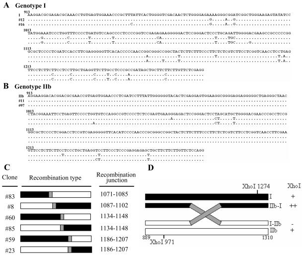
FIG. 2.
HDV-related sequences detected in the serum of a patient with mixed genotype I and IIb infection. (A and B) Alignment of nucleotide sequences (nt 913 to 1280) from representative HDV isolates of genotypes I (A) and IIb (B), and sequences identified in the mixed infection of HDV. Dots indicate conserved nucleotides. Sources of the representative isolates of different genotypes are as follows: I, HDV genotype I isolated from Italy (GenBank accession number M21012) (28); IIb, genotype IIb from the Taiwan-IIb-1 clone (GenBank accession number AF209859) (60). The sequences obtained from two clones each of HDV genotypes I (clones 12 and 16) and IIb (clones 11 and 97) from the mixed infection are shown. (C) Schematic demonstration of HDV recombinants identified by sequence analysis. The recombination junctions are shaded in gray and summarized on the right. The genotype I and IIb sequences are indicated by closed and open bars, respectively. (D) Restriction maps of the potential clones carrying recombinant HDV genomes. The XhoI RFLP profiles and the predicted genome organization of recombinants 5′-I-IIb-3′ and 5′-IIb-I-3′ are summarized on the right.

