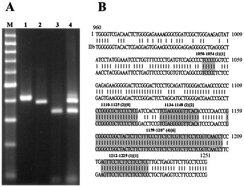
FIG. 5.
HDV recombination in transfected cultured cells. (A) XhoI RFLP analysis of PCR products amplified from total RNA extracted from cotransfected cultured cells by use of consensus primers. Lanes: M, 100-bp-ladder molecular size markers (the dominant band is 500 bp in size); 1, undigested PCR products; 2, digested genotype I PCR products; 3, digested genotype IIb PCR products; 4, a complex RFLP pattern obtained from transfected cultured cells. (B) Primary structure of the crossover regions of intergenotypic HDV recombinants identified in transfected cultured cells by use of genotype-specific primers. The nucleotide sequences of the genomic segments (nt 960 to 1251) of HDV genotypes I (Italian clone) and IIb (Taiwan-IIb-1 clone) are given. Short lines depict the homologous bases between the two genotypes. The crossover regions identified are indicated by gray shading. Numbers given in parentheses and square brackets indicate the number of 5′-I-IIb-3′ and 5′-IIb-I-3′ recombinant clones, respectively.

