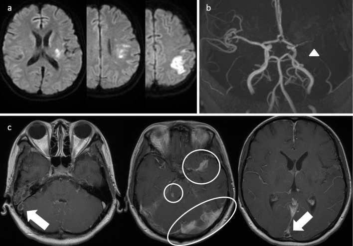
Figure 3.
Cranial MRI findings of recurrent cerebral infarction in tuberculous meningitis. (a) Diffusion-weighted images show infarcted area in the left posterior limb of the internal capsule, left corona radiata, and left parietal lobe. (b) MRA shows occlusion of the left middle cerebral artery (arrow head). (c) Enhanced T1-weighted images show inflammatory changes in the base of the brain (encircled area) and venous sinus thrombosis (arrow). MRI: magnetic resonance imaging, MRA: magnetic resonance angiography

