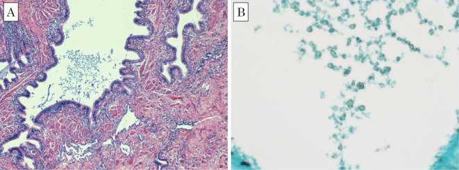
Figure 6.
Light microscopic images of a histopathologic specimen, showing a fungus-like substance in the bronchus. Inflammatory cell infiltration and collagen fiber hyperplasia is noted, but invasion of fungus-like substance into the lung tissue is not definitive. (A) Hematoxylin and Eosin staining. (B) Grocott methenamine silver stain.

