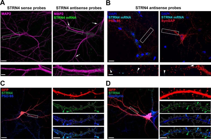
Figure 1.
Strn4 mRNA is dendritically localized, and STRN4 protein is mainly present at excitatory synapses but not inhibitory synapses. A, in situ hybridization was performed on dissociated rat hippocampal neurons (17 DIV) using nucleotide probes targeting STRN4 (STRN4 antisense probes). Discrete Strn4 mRNA puncta (green) were observed in MAP2-positive dendrites (magenta); some of the puncta (arrows) were localized ∼60–80 μm away from the cell body. The green puncta were absent when neurons were hybridized with the sense probes. B, hippocampal neurons (17 DIV) were subjected to Strn4 mRNA in situ hybridization followed by PSD-95 or SynGAP immunofluorescence staining. Some Strn4 mRNA granules (cyan, arrowheads) were in close proximity, but not precisely co-localized, to PSD-95 or SynGAP puncta at excitatory synapses (red). C and D, hippocampal neurons (21 DIV) expressing GFP (red) were co-stained with STRN4 protein (green) and the excitatory postsynaptic protein PSD-95 (blue) or the inhibitory postsynaptic protein gephyrin (blue). Merged images are shown in the bottom right panels, and examples of co-localized puncta are shown (light blue arrowheads). C, co-localization of the STRN4 puncta (green arrowheads) with PSD-95 (blue arrowheads) on dendritic spines. D, many STRN4 puncta (green arrowheads) in the dendritic shaft were distinct from the gephyrin puncta (blue arrowheads). Scale bars, 20 μm (upper panels in A and B and left panels in C and D) or 5 μm (lower panels in A and B and right panels in C and D).

