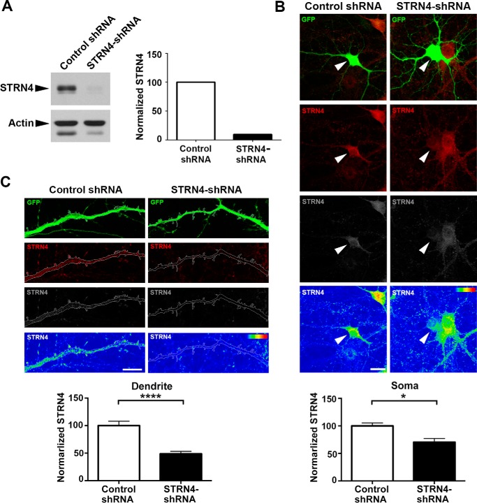
Figure 4.
shRNA-mediated knockdown of STRN4 expression in neuron. A, STRN4 shRNA (or control shRNA) was transfected into rat cortical neurons by nucleofection, and lysate was collected at 5 DIV for Western blot analysis. The intensity of STRN4 was normalized with that of actin. B and C, immunofluorescence staining of STRN4 in hippocampal neurons co-transfected with GFP and the STRN4 shRNA (or control shRNA). Fluorescence intensity of STRN4 (red) was indicated in the heat maps (lower panels). STRN4 immunoreactivity was significantly reduced in both the soma (arrowheads in B) and the dendrites (C) of the GFP-positive, STRN4-shRNA-transfected neurons, as compared with the control shRNA-transfected neurons (eight dendrites from five neurons were quantified for each condition). Data are mean ± S.E.; *, p < 0.05; ****, p < 0.0001; Student's t test. Scale bars, 20 μm (B) and 10 μm (C).

