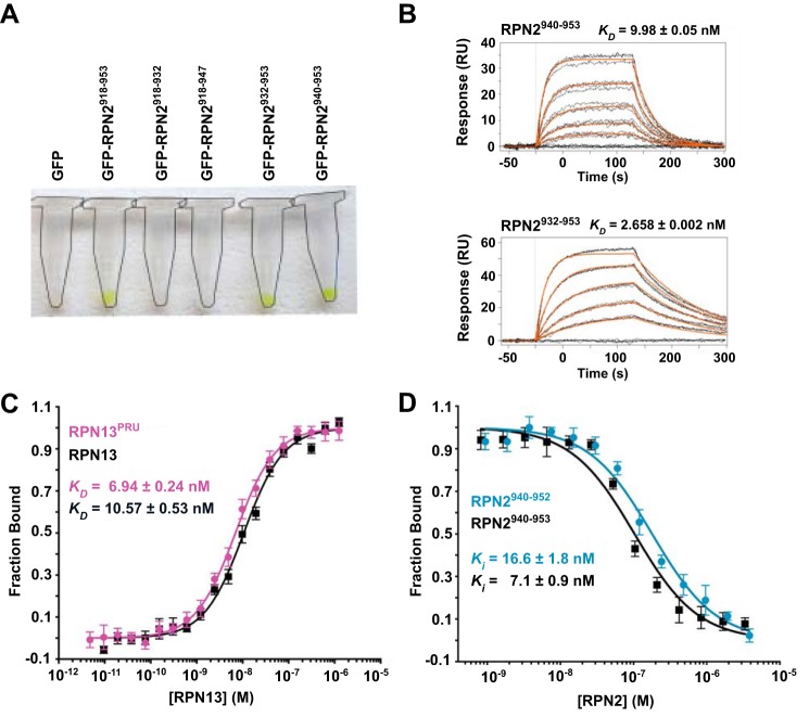
Figure 1.
Examination of the RPN2-RPN13 interaction. A, GFP retention assays used to visualize stable association of various RPN2 peptides with RPN13. Complex formation is visualized by retention of green GFP-RPN2 on the resin. Microcentrifuge tubes outlined for clarity. B, representative SPR sensorgrams illustrating full-length RPN13 binding to RPN2 peptides corresponding to residues 940–953 (top) and 932–953 (bottom). C, representative fluorescence polarization binding curves of full-length RPN13 (black) and RPN13PRU (magenta) binding to fluorescently labeled RPN2(940–952). D, representative fluorescence polarization competition curves of RPN2(940–952) (blue) and RPN2(940–953) (black) competing with fluorescently labeled RPN2(940–952) for binding of RPN13PRU.

