Figure 4.
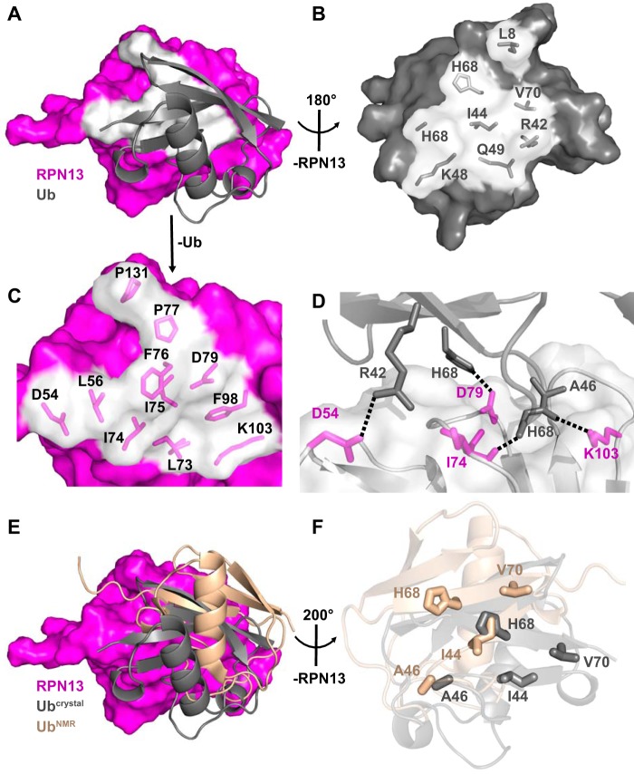
RPN13-ubiquitin interaction. A, overview of the RPN13-ubiquitin interface. RPN13 residues within 4 Å of ubiquitin, white. B, view of ubiquitin residues at RPN13 interface upon ∼180° rotation around y axis of Fig. 3A. RPN13 removed for clarity. C, zoomed view of RPN13 residues at ubiquitin interface as visualized in A. Ubiquitin removed for clarity. D, electrostatic interactions at the RPN13-ubiquitin interface. E, disparity in ubiquitin positioning between crystallographic (gray) and NMR docking (wheat, PDB code 2Z59) models. F, differences in positioning of key residues (shown as sticks) between crystallographic (gray) and NMR docking (wheat) models upon alignment on RPN13.

