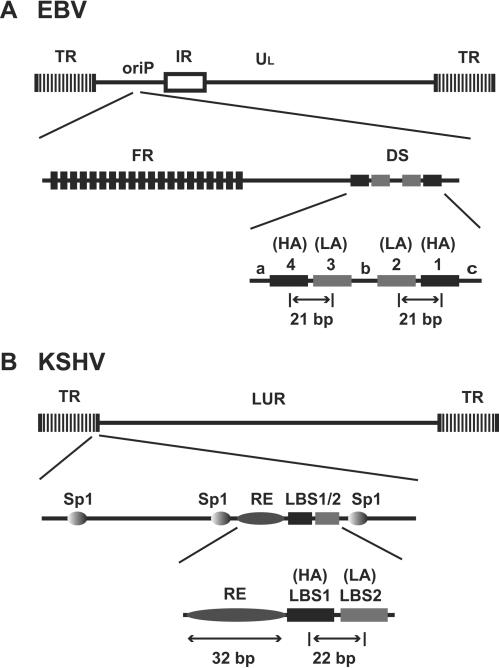
FIG. 4.
Comparison of the latent origins between EBV and KSHV. (A) Genome structure and oriP of EBV. (B) Genome structure and latent origin of KSHV. IR, internal repeat; DS, dyad symmetry; FR, family of repeat; HA, high-affinity binding site; LA, low-affinity binding site; LBS1 and LBS2, LANA binding sites 1 and 2, respectively.

