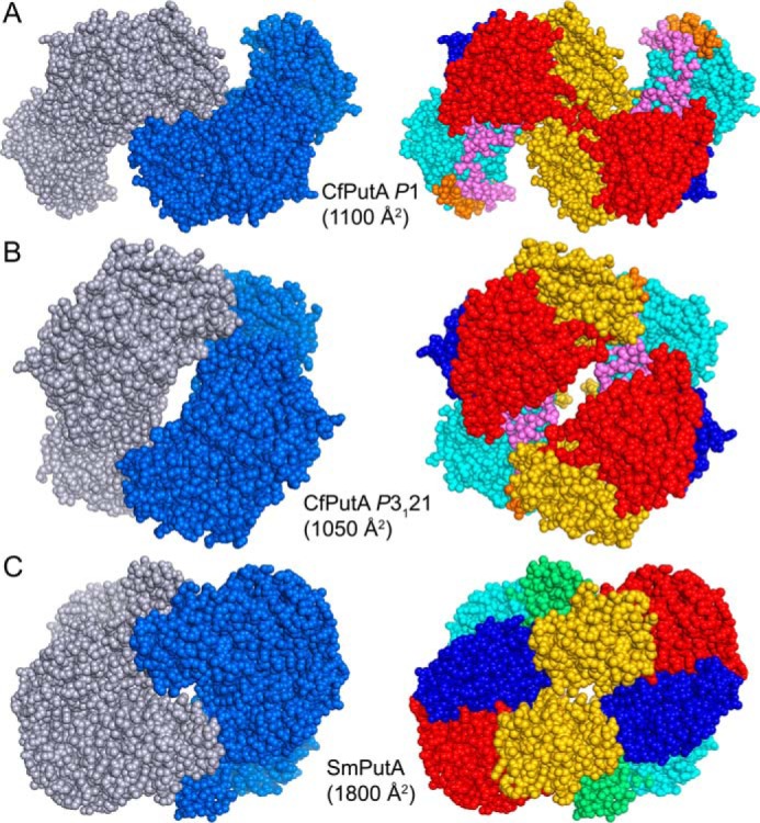
Figure 8.

Dimeric assemblies of CfPutA and SmPutA. A, the largest protein-protein interface in the CfPutA P1 crystal form. B, the largest protein-protein interface in the CfPutA trigonal crystal form. C, the bona fide dimer of SmPutA, which has been validated by SAXS and crystallography (PDB code 5KF6). The interfacial surface areas are listed for each assembly. On the left side, the chains have different colors. On the right side, the domains are colored as in Fig. 4: N-terminal arm (orange), α-domain (green), PRODH barrel (cyan), PRODH-GSALDH linker (violet), NAD+-binding (red), GSALDH catalytic (blue), and C-terminal ALDHSF (gold).
