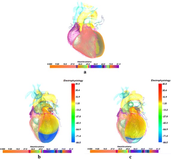
Fig. 5.

Cardiac anatomy structure visualization with traditional light transport model [44] and biophysical merging visualization of electrophysiological excitation conduction in left ventricle at different time with the context of genuine organs of heart (The color mapped by anatomy bar scale and the potential bar scale are not connected but separate). a Cardiac anatomy structure; b 320 ms; c 340 ms
