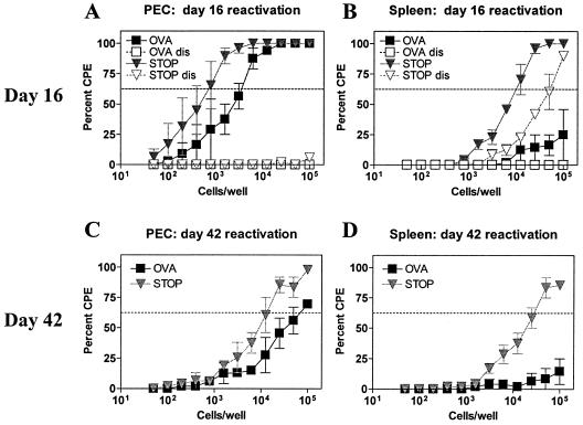
FIG. 6.
OTI T cells control reactivation from latency of γHV68.OVA. Peritoneal cells (PEC) (A) and splenocytes (B) from OTI/B6 mice, infected with 106 PFU, i.p., of either γHV68.OVA (filled squares) or γHV68.v-cyclin.STOP (filled inverted triangles) for 16 days, were evaluated by the ex vivo reactivation assay. Open symbols are samples that were mechanically disrupted and thus represent persistent virus in the samples. Peritoneal cells (C) and splenocytes (D) from OTI/B6 mice were also evaluated at 42 days after infection. No persistent virus was seen in these samples (data not shown). The data are pooled from two independent experiments. CPE, cytopathic effect.

