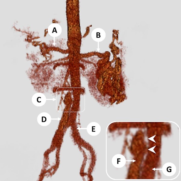
Fig. 1.

Patient 1: 3-D reconstruction images of the aorta from the CT angiogram, demonstrating an abdominal aortic aneurysm with complex dissection extending into the right common iliac artery. A, common hepatic artery; B, splenic artery (tortuous); C, top of the aortic aneurysm and dissection (magnified in inset); D, false lumen extending into the right common iliac artery; E, true lumen extending into the left common iliac artery; F, false lumen of the aorta; G, true lumen of the aorta. Inset Arrowheads indicate dissection flap in the aorta.
