
Fig. 5.
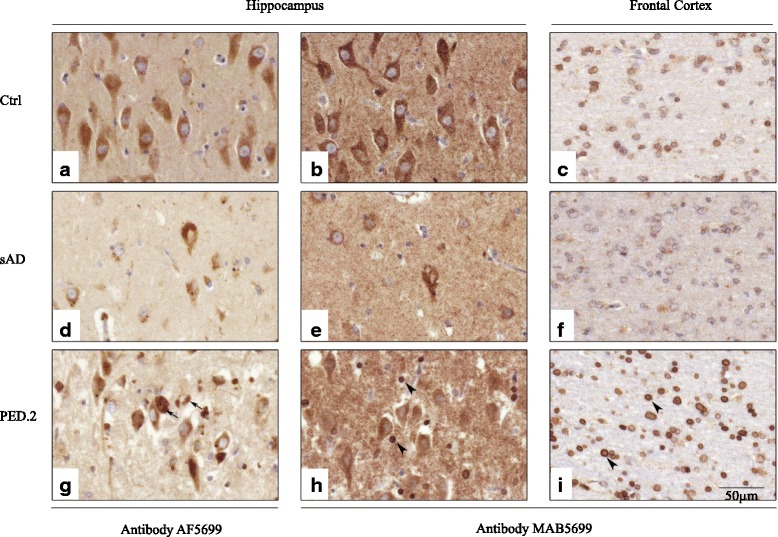
Immunohistochemical localization of SORL1 in postmortem brain material from controls, sporadic AD and PED.25. Representative pictures from control (a-c), sporadic AD (d-f) and PED.25 (g-i) in the CA1 region of hippocampus (a-b, d-e, g-h) and subcortical white matter in frontal cortex (c, f, i) using two different SORL1 antibodies, AF5699 (a, d, g) and MAB5699 (b-c, e-f, h-i). The AF5699 SORL1 antibody showed an intense immunoreactivity of extracellular SORL1 aggregates in PED.25 (arrows in g). Arrowheads indicate strong SORL1 immunoreactivity (MAB5699) in glial cells in grey (h) and white matter (i) in the affected member from PED.25. Scalebar: 50 μm. Ctrl = control, sAD = sporadic AD, PED.25 = affected family member II:6