. 2017 Mar 6;8(5):3703–3711. doi: 10.1039/c6sc04897h

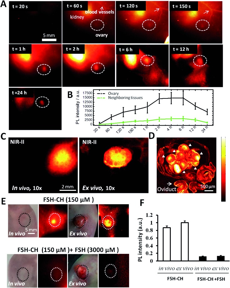
Fig. 2. NIR-II imaging of ovarian follicles using follicle stimulating hormone-fluorophore CH1055 (FSH-CH) in adult female mice: time-course and specificity. (A) FSH-CH (12.5 μg) was injected into the tail vein of an adult female mouse before NIR-II imaging at different post-injection times. There was a rapid accumulation of signals in the blood vessels and kidney at 60 s. Ovarian signals begin to show up at 120 s and peak at 2–6 h after injection, showing a sustained retention for up to 24 h. Side view images are shown to focus on one ovary. (B) Quantitation of the fluorescence intensity in the ovary at different time points (n = 3). (C) Imaging at high magnification at 24 h after FSH-CH injection. Ovarian NIR-II signals inside the body (in vivo) and after ovary exposure from the abdominal cavity with the uterus, vasculature and nerves connected (ex vivo) are shown. (D) Confocal image of the same ovary showing bright signals in granulosa cells inside individual follicles (arrowheads) together with background signals in the oviduct (arrow). (E) Displacement of FSH-CH binding by non-conjugated FSH in the ovary. Adult female mice were injected with FSH-CH (12.5 μg of FSH) or FSH-CH plus a 20-fold excess of unconjugated FSH (250 μg) before imaging 2 h later. Strong NIR-II signals were found in the ovary both in vivo and ex vivo whereas no signal was found in the ovary when excess FSH was injected together with FSH-CH. Light microscope pictures accompany each NIR-II image. (F) Quantitation of NIR-II signals in individual groups. Error bars indicate the standard deviation of each group. PL, photoluminescence.

Fig. 2