FIG. 1.
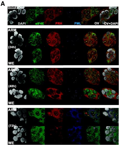
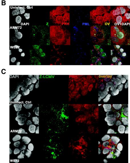
Confocal images of intracellular localization of PRH. Hepatic HepG2 or Huh7 cells were grown on coverslips and infected with LCMV as described in Materials and Methods. (A) Infected or uninfected HepG2 cells were stained with anti-PRH (red), anti-PML (blue), or anti-eIF4E (green) to determine how infection affects the localization of PML, PRH, and eIF4E in hepatic cells. Huh7 cells (B) or HepG2 cells (C) were stained with anti-PRH (red), anti-PML (blue), or anti-LCMV Z (green). All cells were counterstained for DNA with DAPI (gray). The overlay is shown in yellow (OV), and the overlay of DAPI staining is designated OV+DAPI. These confocal images represent single optical slices through the cells. Magnification, ×300. FITC and Texas Red channels were recorded independently.


