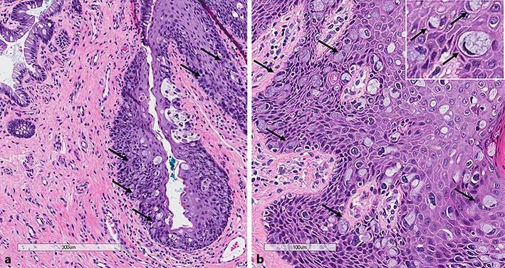
Fig. 1.

Hematoxylin and eosin stains show intraepithelial spread of Paget cells involving the anal transitional zone. a The squamo-columnar junction is seen. b At the upper right corner (inset, ×40) Paget cells with large cytoplasmic mucin globules and peripherally displaced nuclei are shown.
