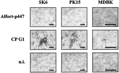
FIG. 3.
Growth properties of CP G1 in different cell lines. Porcine SK6 (left) and PK15 cells (middle) as well as bovine MDBK cells (right) were infected with noncp CSFV Alfort-p447 (top) or CP G1 from the first cell culture passage (middle). Noninfected (n.i.) cells (bottom) served as negative controls. After infection, only porcine cells were overlaid with medium containing 0.6% low-melting-point agarose. After an incubation period of 66 h at 37°C, a CPE was only observed after infection with CSFV CP G1. Scale bar, 200 μm.

