
Fig. 1.
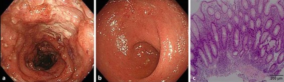
Colonoscopy findings before treatment: observation up to the transverse colon. Extensive undermining ulcers, severe mucosal edema, and scope contact-induced hemorrhage were observed in the transverse colon (a) over the sigmoid colon. Although mucosal edema was present in the rectum (b), only partially visible vascular patterns and erosion were observed. Results of rectal biopsy (c): mucosa with relatively marked inflammatory cell infiltration including neutrophils and regenerative changes. No epithelioid granuloma or active vasculitis was observed. The histopathological diagnosis was ulcerative colitis with mild activity, compatible ulcerative colitis. Diffuse inflammatory cell infiltration (++), reduction of germinal (goblet suspicion) cells (+), regressive change of glandular epithelium (±), crypt abscess (–), disturbed gland duct arrangement (+), gland regeneration (+), Paneth cells (–).