
Fig. 4.
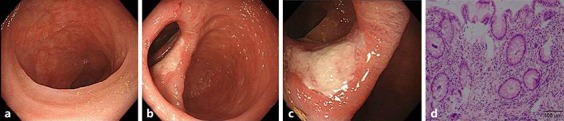
Colonoscopy findings at week 6 of tacrolimus administration. The scope was inserted up to the end of the ileum. No reddening or erosion was observed at the end of the ileum (a). Bauhin's valve (b) was slightly dilated, and a round ulcer was formed in the inferior lip of Bauhin's valve. In the ileocecum over the ascending colon, visible vascular patterns were reduced. In the transverse colon (c) over the sigmoid colon, ulcer scars and inflammatory polyps were noted. Visible vascular patterns were partially improved. Rectal erosion had resolved as observed on the previous colonoscopies. Results of biopsy from the margin of the ulcer in Bauhin's valve (d): the mucosa showed mild activity. Gland tissue showed extension and slight dentate changes, but the nuclei were small. Diffuse inflammatory cell infiltration (+), decrease in germinal (goblet suspicion) cells (±), regressive change of glandular epithelium (–), crypt abscess (–), disturbed gland duct arrangement (+), gland regeneration (+), Paneth cells (–).