
Fig. 1.
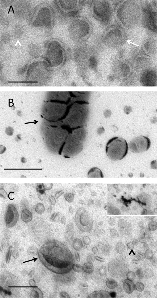
Liposomes. Liposomes in methylcellulose films containing uranyl acetate (A), phosphotungstic acid (B) and uranyl acetate/phosphotungstic acid mixture (C). Note the difference between the clarity of display of the membrane structures visualized with UA and UA/PTA (A and C respectively; larger structures, arrows and smaller structures, arrowheads). Inset in C shows characteristic precipitates generated in the mixed UA/PTA stain. The clumping of vesicles that occurs with PTA (arrow in B) alone is not apparent when the UA/PTA mixture is used. Bars 100 nm.