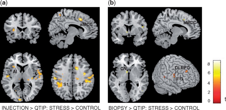
Fig. 1.
Activation differences for the group × condition contrasts: (a) INJECTION > QTIP: STRESS > CONTROL, showing activation in left anterior insula (AI), anterior midcingulate cortex (aMCC) and bilateral somatosensory cortex (S1) (b) BIOPSY > QTIP: STRESS > CONTROL, showing activation in anterior midcingulate cortex (aMCC), left caudate nucleus (CN), right inferior frontal gyrus (IFG) and right DLPFC. Correction for multiple comparisons was performed using voxel-level family-wise error correction at P < 0.05 over the whole brain.

