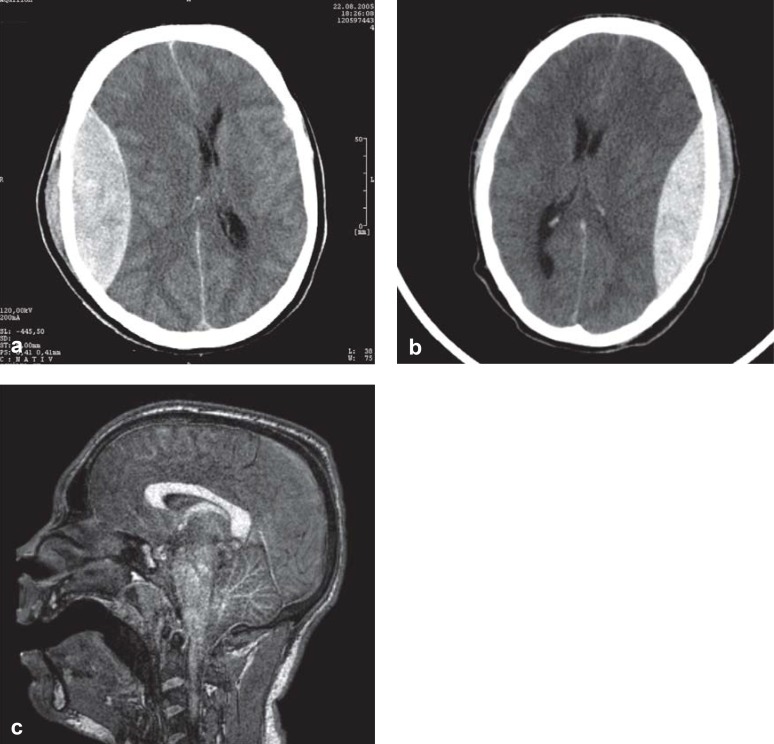
Figure 1.
Head CT and MRI in patients with traumatic brain injury.
This 42-year-old woman (physician) was comatose 3 hours after an accident, with acute pathological extensor responses. The CT shows an epidural hematoma. She awakened promptly after surgical removal of the hematoma.
This 20-year-old man fell, remained mentally lucid, and went home; a few hours later, he was found unconscious in bed with fixed and dilated pupils. The CT shows an epidural hematoma, which was removed immediately, like the one in (a).
After surgery, he remained comatose and his pupils remained fixed and dilated. The MRI shows increased signal intensity throughout the brainstem, evidently reflecting a pathological abnormality that arose secondarily, after the lucid interval, as a result of sustained pressure on the brainstem arising from the epidural hematoma. The superiority of MRI to CT in this case is clear, as the lethal brainstem injury cannot be seen in the CT in (b), just as the CT in (a) cannot show the absence of a brainstem lesion.

