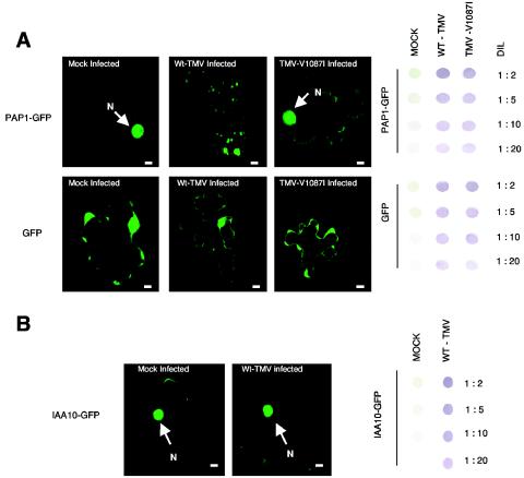
FIG. 6.
Transient expression of PAP1-GFP in N. benthamiana leaf tissues. (A) Fluorescent images of cells expressing a PAP1-GFP fusion protein or GFP alone in noninfected (mock-infected), WT-TMV-infected, or TMV-V1087I-infected tissue. Bars, 10 μm. Immunodot blots showing dilutions (DIL) of leaf tissue homogenate used to monitor virus levels in PAP1-GFP-transformed leaf tissues are shown to the right of the fluorescent cell images. (B) Fluorescent cell images of IAA10-GFP in either mock- or TMV-infected tissue.

