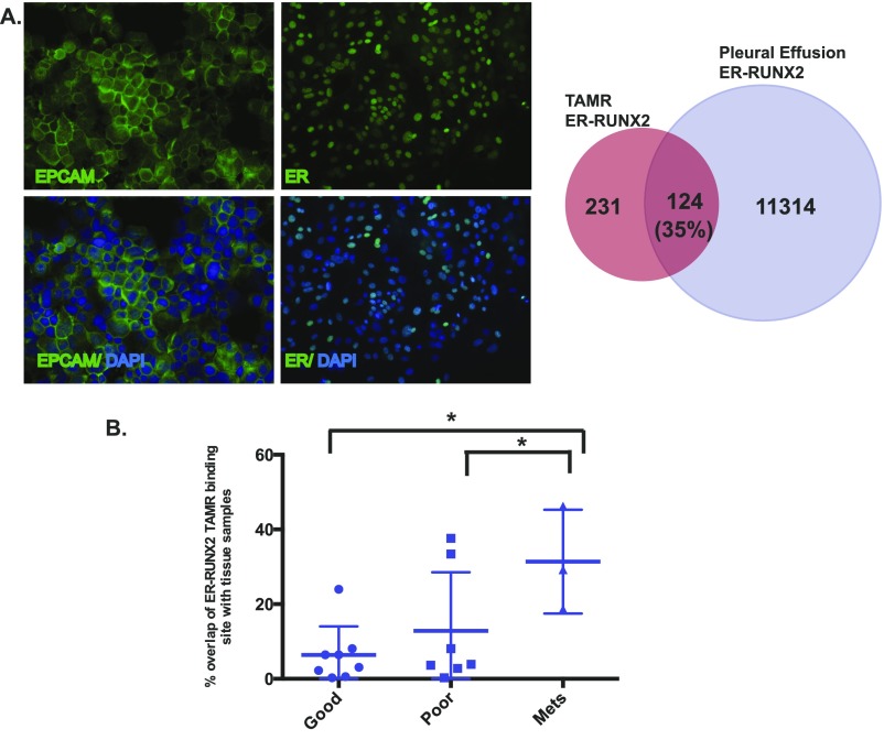
Fig. S4.
(A) Immunofluorescence staining of cells isolated from a malignant pleural effusion. Staining is for epcam and DAPI (Left) and ERα and DAPI (Right). Magnification, 20×. Venn diagram showing the overlap between the ER–RUNX2 binding sites enriched in the TAMR cells and the pleural effusion metastatic cells in ER binding sites with a RUNX motif within ±1 kb from the summit. (B) Comparison of overlap between the ERα–RUNX2 binding sites in TAMR cells with published ERα binding in ER+ breast cancer tissue samples including primary breast cancers of good prognosis and poor prognosis and metastatic samples. Unpaired Student’s t test was used to calculate the P values. *P < 0.05.

